Ophthalmology
If your pet is suffering from damage to its eyes or is having trouble seeing you should make an appointment with our ophthalmology service as soon as possible.
Below are the services offered at Canary:
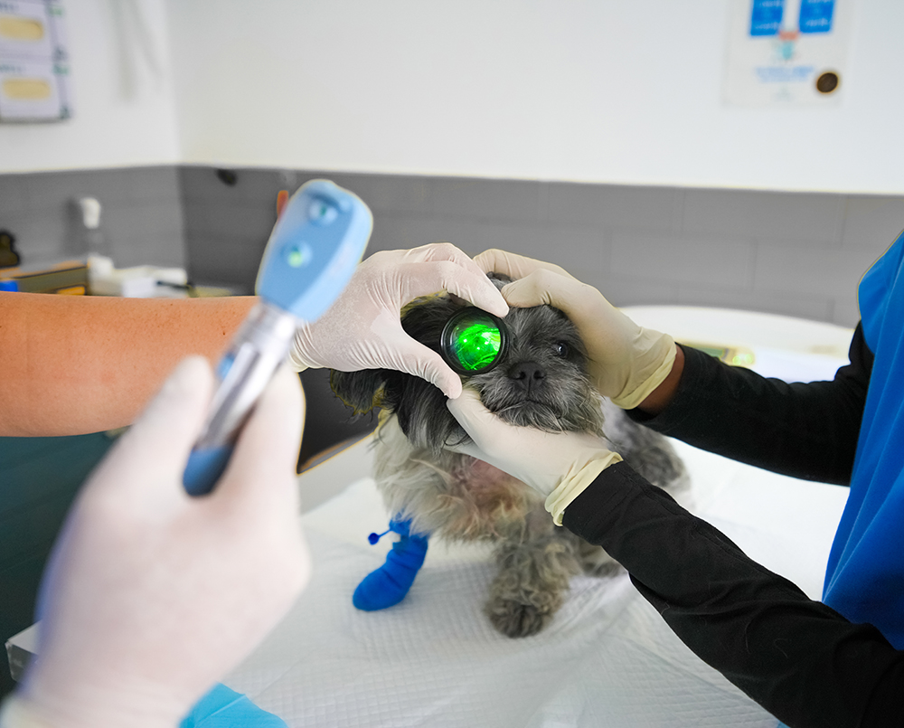
- General eye examination with direct ophthalmoscopy, stains, Schirmer tear test and so on to make sure the eyes of your animal are ok.
- Tonometry is a test used to evaluate a patient for glaucoma and to monitor the progression of glaucoma. It is performed with an instrument called a Tono-Pen, a hand held instrument used to measure fluid pressure inside the eye, or intraocular pressure (IOP). By gently tapping on the cornea three measurements are taken. The unit then displays an average of the readings.The fluid inside the eye is called the aqueous humor. The aqueous humor is produced and drains from the eye at approximately the same rate, resulting in a stable pressure inside the eye of 15 to 20 mmHg (millimeters of mercury). Glaucoma occurs as a consequence of a defect in the drainage angle resulting in inadequate outflow of aqueous humor and a subsequent buildup of pressure inside the eye. The resulting high pressure damages the optic nerve and results in blindness.
- Eyelid surgery is used to correct abnormalities of the eyelid in dogs and cats. Eyelid abnormalities can occur for many reasons. The most common causes of eyelid abnormalities that need surgery include; entropion, ectropion, distichaisis, ectopic cilia, eyelid tumors, cherry eye, eyelid laceration, proptosis and eyelid agenesis. Many of these conditions if left untreated will cause discomfort due to corneal ulcerations. Eyelid surgery is best done by a veterinary ophthalmologist as this type of surgery is plastic surgery and takes years of training to master.
- Corneal surgery is used to repair abnormalities of the surface of the eye. The cornea is the wind shield of the eye and is vital for vision to keep clear. The most common causes of the corneal abnormalities that require surgery include; corneal ulcerations, corneal perforation, corneal edema, corneal lacerations, corneal squestrum, corneal/limbal tumor and corneal dermoids. Surgeries to repair or correct these conditions are performed under an operating microscope and often require a covering or graft to help the wound heal. The most common graft is to use the patients own conjunctiva, the lining of the eyelids.
- Ocular ultrasound is a noninvasive diagnostic modality to evaluate for ocular abnormalities and can be used as a complement to traditional ophthalmoscopic examination.This ultrasound based diagnostic medical imaging technique is used to evaluate the eye structure and any pathological conditions in real-time images. Veterinary Ophthalmology services treat complicated or difficult problems such as cataracts, corneal ulcers, Entropion, Glaucoma, prolapsed gland of the nictitans (cherry eye) and Uveitis.


